Imaging Technique Reveals DNA’s Hidden Shape in the Earliest of Embryos

After fertilization, the gametes within the egg prepare to coordinate the formation of a new life. Genomicists have traditionally thought that this period, before a coordinating process called zygotic genome activation begins, is marked by genome disorganization. This initial period, they believed, was just a liminal transition state before the embryo began to sort itself out and start making a complex new organism.
Now, a pair of new studies suggests that, in this earliest stage of life, the genome is actually more organized than we first thought.
Read More: A Lab Has Created a Synthetic Human Embryo Without a Fertilized Egg
What is Pico-C?
In the first study, published in Nature Genetics, scientists have unveiled a tool called Pico-C that reveals the 3D structure of the genome during the first days of life. The tools reveal how this structure, called chromatin, is organized.
Pico-C showed that before zygotic genome activation begins, the genome has already packaged itself into a structure. The findings could help us understand these key steps in development, which can cause serious and lifelong health problems when they go wrong.
“We used to think of the time before the genome awakens as a period of chaos,” said Noura Maziak, an epigenomicist at Imperial College London and co-author of the new study, in a press release. “But by zooming in closer than ever before, we can see that it’s actually a highly disciplined construction site. The scaffolding of the genome is being erected in a precise, modular way, long before the ‘on’ switch is fully flipped.”
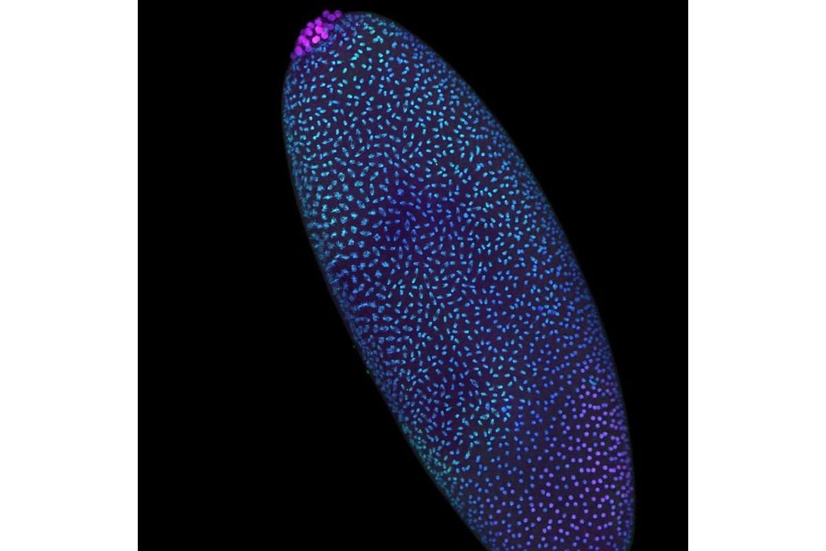
Fruit Fly DNA Structure
The team made their findings using Pico-C to image fruit fly (Drosophila) embryos. The fly’s rapid developmental timeline, in which thousands of cells are formed in just a few hours of nuclear divisions, makes it an ideal model for developmental biologists.
When they looked at the fly’s developing genome, they saw an ordered structure, rather than a messy maze. The fly’s DNA was folded into loops, which allowed external regulators to alter how the genome developed.
The DNA’s props and actors were being carefully arranged on the stage, ready for showtime. The team even identified so-called “pioneer” factors that, when depleted, disrupted this careful early organization.
Pico-C is set to help us understand more than fruit fly development. In a second study, led by researchers at ETH Zürich in Switzerland and sharing some of the same authors as the Nature Genetics paper, the tool was tested in human cells. This second paper was published in Nature Cell Biology.
Here, the researchers tested the effects of removing chromatin “tethers” that maintain the genome’s structure. They saw that disrupting the genome’s architecture triggered a dramatic turn of events: the cell misinterpreted the collapse as a viral attack, setting off a wave of inappropriate autoimmune attack. This type of misdirected activation could lead to disease.
“These two studies tell a complete story,” said Juanma Vaquerizas, a developmental epigenomicist at Imperial College London who co-authored both studies, in the press release. “The first shows us how the genome’s 3D structure is carefully built at the start of life. The second shows us the disastrous consequences for human health if that structure is allowed to collapse.”
These studies could be just the first example of how Pico-C could help us understand the genome’s development. Importantly, the tool requires only a tiny sample for analysis, ten times less than other procedures require. The researchers hope this could help Pico-C reveal how the changing shape of DNA controls gene regulation in health and disease.
Read More: More Like a Movie Than a Portrait — A New DNA Model Redefines Family History
Article Sources
Our writers at Discovermagazine.com use peer-reviewed studies and high-quality sources for our articles, and our editors review for scientific accuracy and editorial standards. Review the sources used below for this article: