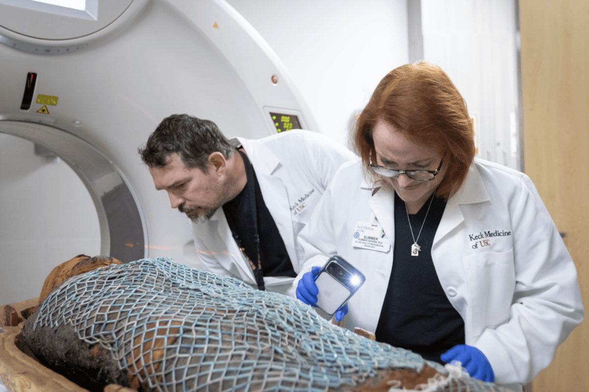
It’s not every day that you get to run a mummy-filled sarcophagus through a CT scanner. But a research team at the University of Southern California (USC) has been doing just that to help peel back the linen-wrapped past of ancient Egypt.
Using hospital-grade CT scanners, the team from Keck Medicine of USC has examined two ancient Egyptian mummies, offering an unusually intimate look at how these men lived, aged, and experienced pain more than 2,000 years ago.
What the scans revealed goes far beyond what researchers expected and humanized the mummies in strikingly modern ways.
“[One of the mummies] had a hip issue that would definitely have been visible. This would have been a disability for him. So it’s just really interesting to relate to that because modern humans have these back issues and hip issues […] and that makes us understand what he went through,” says Summer Decker, Ph.D., 3D imaging lead for Keck Medicine of USC and director of the USC Center for Innovation in Medical Visualization.
3D digital model of Nes-Min.
(Image Courtesy of Keck Medicine of USC)
What the Scans Revealed About Ancient Egyptian Lives
Radiologists conducted full-body CT scans of two Egyptian priests: Nes-Min, who lived around 330 B.C.E., and Nes-Hor, from roughly 190 B.C.E. High-resolution, 320-slice CT imaging exposed fine anatomical details, including eyelids, lips, and bone wear.
As Decker explains, the energy in the room during the scans was filled with excitement and awe: “We had folks from the California Science Center there and they had visions of the kind of imaging that had been done in the past. But it’s hard to explain what a 325 CT scanner can do. So when they saw the models coming off, they could immediately see the eyelids and the eyes and really fine details in the mummies’ bodies. I remember a moment when their mouths were hanging open, and I had to keep reminding them that this is what we do every day for our patients.”
The older mummy, Nes-Min, showed signs of a collapsed lumbar vertebra, suggesting chronic lower back pain consistent with natural aging and physical strain. He was also buried with scarab beetles and fish-shaped artifacts that the research team was able to recreate using 3D printing.
“I was showing some people the scarabs that we found. We 3D printed them and they are high resolution, one-to-one replicas,” explains Decker. “This is the first time in over 2,000 years that someone’s held them.”
Nes-Hor, meanwhile, appeared to have lived longer than Nes-Min but endured significant physical hardship. His scans revealed dental disease and a severely deteriorated hip that would have been visible and disabling during his lifetime.
Read More: A 1,000-Year-Old Mesoamerican Mummy Reveals an Ancient Man’s Microbiome
How CT Scanning and 3D Printing Brought Mummies to Life
Nes-Hor’s skull and facial structure as seen in a 3D model.
(Image Courtesy of Keck Medicine of USC)
The technology behind these discoveries is the same used daily in hospitals. CT scanners generate hundreds of cross-sectional “slices” of the body, which are digitally stacked to create detailed 3D models. In this case, those models were not only analyzed on screen but also physically reproduced using 3D printers.
These prints are safe to handle and are precise down to the millimeter, allowing researchers and the general public a chance at an up-close-and-personal look at a piece of history without destroying the original specimens.
Where Can You See the Mummies and 3D Printings?
Decker and her team are actively working on the analysis and reconstructions, so much so that she was in the middle of a printing project while Discover spoke with her: “We’re working on it right now. We’re 3D printing Nes-Min’s ribs because he had healed fractures probably from an injury from when he was much younger. They healed in a kind of dislocated way.”
Beyond the in-lab discoveries, the mummies, scans, and 3D prints will be on display during the “Mummies of the World: Exhibition,” opening February 7 at the California Science Center.
“All of this was an effort through collaboration,” concludes Decker. “These kinds of scientific advancements happen when people work together, not just in a silo. Then to be able to display that so the general public can see is a great example of public science.”
Read More: The First-Ever Whole Genome of an Ancient Egyptian Reveals What Life Was Like 4,800 Years Ago
Article Sources
Our writers at Discovermagazine.com use peer-reviewed studies and high-quality sources for our articles, and our editors review for scientific accuracy and editorial standards. Review the sources used below for this article: